
Cell Division:
Understanding the Fundamental Process
Cell division is a fundamental process in biology, essential for the growth, development, and repair of multicellular organisms. It involves the replication and distribution of genetic material to ensure each daughter cell receives a complete set of chromosomes. This intricate process occurs in a series of well-defined phases, each crucial for the accurate transmission of genetic information. Understanding these phases provides insights into the complexity and precision of cellular replication.
-
Interphase: Preparing for Division
Interphase is the preparatory phase preceding cell division. Despite being a distinct phase, it’s often not included in the typical breakdown of the cell cycle. Interphase can be further divided into three stages:
1.1 G1 Phase (Gap 1):
During this phase, the cell grows, synthesizes proteins, and carries out its normal functions. It also monitors its environment to ensure conditions are good for division. Critical checkpoints exist here to regulate progression to the next phase.
1.2 S Phase (Synthesis):
In this phase, DNA replication occurs. Each chromosome is duplicated, resulting in sister chromatids held together by a centromere. The process ensures that each daughter cell will have a complete set of genetic material.
1.3 G2 Phase (Gap 2):
Following DNA replication, the cell continues to grow and prepare for division. Additional organelles are synthesized, and the cell undergoes further checks to ensure DNA integrity and proper replication. Once again, checkpoints monitor the cell’s readiness for division.
-
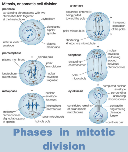
Mitotic Phase: Dividing the Cell
The mitotic phase encompasses the actual division of the cell’s nucleus and cytoplasm. It consists of several distinct stages:
2.1 Prophase:
Chromatin condenses into visible chromosomes, each consisting of two sister chromatids. The nuclear envelope disintegrates, and the mitotic spindle begins to form, composed of microtubules emanating from centrosomes located at opposite poles of the cell.
2.2 Prometaphase:
The nuclear envelope is fully disassembled, allowing microtubules to interact with the chromosomes. Kinetochores, protein structures on the centromeres, attach to spindle fibers, facilitating chromosome movement.
2.3 Metaphase:
Chromosomes align along the metaphase plate, an imaginary plane equidistant between the two poles of the cell. This alignment ensures that each daughter cell will receive an identical set of chromosomes during division.
2.4 Anaphase:
Sister chromatids separate and move toward opposite poles of the cell, pulled by the shortening microtubules of the spindle apparatus. Once separated, each chromatid is considered a distinct chromosome.
2.5 Telophase:
Chromosomes arrive at opposite poles of the cell and de-condense back into chromatin. A new nuclear envelope forms around each set of chromosomes, and the spindle apparatus disassembles. Meanwhile, cytokinesis, the division of the cytoplasm, begins.
-
Cytokinesis: Completing the Division
Cytokinesis is the final stage of cell division, during which the cytoplasm is divided between the two daughter cells. In animal cells, a contractile ring composed of actin filaments forms at the cell’s equator, pinching the cell membrane inward until the cell is bisected. In plant cells, a new cell wall known as the cell plate forms at the equator, eventually dividing the cell into two distinct daughter cells.
Conclusion:
Cell division is a meticulously regulated process essential for the growth, development, and maintenance of organisms. The coordinated progression through interphase, mitosis, and cytokinesis ensures the accurate transmission of genetic material to daughter cells, preserving genetic integrity and facilitating the perpetuation of life. Understanding the intricacies of cell division provides insight into fundamental biological processes and their regulation.